Digital radiography with Dexis represents a modern approach to dental imaging that places clarity and patient safety at the center of diagnosis. Instead of traditional film, Dexis uses compact electronic sensors to capture high-resolution images of teeth, roots, and surrounding bone. These images are immediately available on-screen, which speeds up the diagnostic process and helps your dental team make informed decisions during the same visit.
Because Dexis sensors produce detailed images, subtle changes in tooth structure and bone density become easier to spot. That means early-stage problems — such as small cavities between teeth or the beginnings of bone loss — are more likely to be detected before they progress. The system’s image quality also supports precise treatment planning for restorative work, endodontic care, and periodontal evaluation.
For patients, the shift to digital radiography is largely invisible but meaningful: there’s no chemical processing, fewer repeat exposures, and the ability to review and discuss findings with your clinician in real time. The result is a smoother visit, clearer communication, and imaging that directly supports high-quality, conservative dental care.
One of the biggest practical advantages of the Dexis platform is speed. Images appear on the monitor seconds after acquisition, allowing clinicians to assess conditions without delay. This immediacy reduces appointment time and eliminates the uncertainty that sometimes accompanies film-based workflows. When an issue is identified, the team can move quickly from observation to recommended next steps.
Beyond speed, Dexis offers software features that enhance interpretation: adjustable contrast, zoom, measurement tools, and comparative viewing modes. These tools help clinicians evaluate lesion size, root canal anatomy, restorative margins, and bone levels with greater confidence. The clarity of digital images can improve diagnostic accuracy, which in turn supports better long-term outcomes for patients.
Because images can be displayed and annotated during your appointment, clinicians can walk patients through what they see and why a particular treatment is advised. That shared visual reference often makes it easier for patients to understand the rationale for care and to participate in treatment decisions with clearer expectations.
Dexis digital radiography significantly reduces radiation exposure compared with conventional film x-rays. The sensitive digital sensors require less energy to produce a diagnostic image, and advanced image processing compensates for lower doses. While all radiographic imaging is used judiciously, this dose reduction is an important safety benefit for routine exams and follow-up imaging.
Digital images also eliminate the need for chemical development, which means fewer hazardous materials in the office environment. From an environmental and safety standpoint, moving away from film-processing chemicals is a welcome improvement that aligns with modern clinical best practices.
Once captured, radiographs are stored securely in the patient’s digital record, making it easier to track changes over time and to maintain complete, organized files. When coordination with a specialist or another practice is necessary — with the patient’s permission — images can be shared quickly and securely to support collaborative treatment planning and continuity of care.
Dexis sensors are designed with patient comfort in mind. They are generally smaller and thinner than film holders of the past, which reduces gagging and discomfort during placement. The quick capture time minimizes how long a sensor needs to remain in the mouth, which helps make intraoral imaging less stressful for patients of all ages.
From the provider perspective, the ergonomic design and instant feedback streamline clinical workflows. Practitioners can retake an image immediately if positioning needs adjustment, rather than waiting for film processing. This reduces the chance of missed anatomy or unclear images and cuts down on repeated exposures.
The ability to show patients their own x-rays on a monitor is a powerful educational tool. Clinicians can point out areas of concern, compare past and present images, and use visual evidence to explain treatment options in plain language. That transparency supports informed decision-making and builds trust between patients and their care team.
At Park Dental Wellness, Dexis digital radiography is integrated into routine exams and diagnostic workflows to support comprehensive, patient-centered care. We rely on high-quality imaging for everything from preventive assessments to more complex restorative and periodontal planning. The technology complements other advanced systems in our office and helps ensure treatments are based on accurate, up-to-date information.
When you come in for an exam, our team uses Dexis images to document current oral health and to establish a baseline for future comparisons. These records are reviewed with you during the visit so you can see precisely what the clinician observes and why a particular follow-up or procedure may be recommended. That collaborative approach helps patients understand their oral health and pursue conservative, evidence-based care.
Dexis also supports interdisciplinary coordination when care involves specialists. With patient consent, images and notes can be shared securely to facilitate referrals and collaborative treatment planning. This interoperability helps maintain continuity of care and reduces delays in diagnosis or treatment.
In short, digital radiography with Dexis is a practical, patient-friendly technology that enhances diagnostic accuracy, reduces exposure, and improves how our team communicates findings and plans care. If you’d like to learn more about how we use advanced imaging at Park Dental Wellness or have questions about what to expect during an exam, please contact us for more information.


Dexis digital radiography is a modern intraoral imaging system that uses compact electronic sensors to capture high-resolution images of teeth, roots and surrounding bone. Unlike traditional film which requires chemical processing, Dexis produces images instantly on a monitor so clinicians can review results during the same appointment. The digital workflow reduces repeat exposures and eliminates film development, making imaging faster and more efficient.
The system includes software tools for contrast adjustment, zooming and measurement that support more detailed interpretation than static film. Those features help clinicians detect subtle changes in tooth structure and bone density earlier than might be possible with film. Overall, Dexis emphasizes clearer images, quicker diagnostics and streamlined record keeping.
Dexis improves diagnostic accuracy by producing high-resolution images that make small lesions and early bone changes easier to detect. Adjustable image settings and measurement tools allow clinicians to evaluate restorative margins, root anatomy and lesion size with greater precision. The ability to compare current and prior images side by side also helps track subtle changes over time.
Instant feedback reduces uncertainty and the need for repeat exposures due to improper positioning or processing errors. When clinicians can confirm findings immediately, they can develop a treatment plan with more confidence and discuss options with the patient while the information is fresh. This leads to more targeted, conservative care based on reliable imaging.
Yes. Dexis sensors are more sensitive than traditional film, which means they require less x-ray energy to produce a diagnostic image. Advanced image processing compensates for lower doses while still delivering clear images suitable for clinical decision making. Reduced exposure is an important safety advantage for routine exams and follow-up imaging.
Clinicians still follow the principle of ALARA, keeping exposures as low as reasonably achievable and ordering radiographs only when clinically indicated. Sensor design and faster capture times also minimize the need for retakes, further limiting cumulative exposure. Together, these practices help maintain patient safety while supporting accurate diagnosis.
Images captured with Dexis are stored digitally in the patient record, which simplifies tracking changes and maintaining complete, organized files. Digital storage eliminates physical film archives and allows clinicians to retrieve past images quickly for comparison or review. Secure practice management systems protect access to these records in accordance with privacy standards.
When coordination with a specialist or another office is necessary, images can be shared electronically with patient consent to support collaborative treatment planning. Secure transfer protocols and encrypted networks help preserve confidentiality during transmission. This interoperability reduces delays and improves continuity of care when multiple providers are involved.
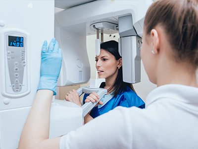
During an appointment, a small Dexis sensor is positioned inside the mouth to capture the required views, and the image appears on the monitor within seconds. The process is quick and usually requires only a brief moment with the sensor in place, which helps reduce discomfort and gag reflex in many patients. If an image needs repositioning, the clinician can retake it immediately without waiting for film processing.
Clinicians often review and annotate images in real time, walking patients through what they see and explaining recommended next steps using the visual reference. That immediate discussion helps patients understand their oral health and participate in treatment decisions. Overall, the appointment tends to be more efficient and educational than workflows based on film.
Dexis sensors are generally thinner and smaller than older film holders, which can reduce discomfort and minimize gagging during intraoral placement. The short capture time means sensors need to remain in the mouth for only a few seconds, making the experience less stressful for children and sensitive patients. Positioning aids and gentle technique further improve tolerance for imaging procedures.
For particularly anxious patients, clinicians can modify their approach and use distraction or positioning strategies to increase comfort. The ability to take an accurate image quickly reduces the likelihood of repeat exposures, which benefits both comfort and safety. Pediatric imaging protocols are also tailored to minimize exposure while capturing diagnostic views.
High-resolution Dexis images reveal fine details of tooth anatomy, restorative margins and root canal systems that are essential for precise treatment planning. Clinicians can measure lesion size, assess bone levels and confirm the fit of restorations with the software tools available in the Dexis platform. This level of information supports predictable outcomes for crowns, fillings, root canals and other restorative procedures.
Immediate image review allows the care team to adapt treatment recommendations on the spot and to discuss options with the patient using visual evidence. Better visualization reduces surprises during operative work and helps prioritize conservative approaches when appropriate. When specialist input is needed, clear digital images facilitate coordinated planning and referrals.
Frequency of dental x-rays depends on individual risk factors, oral health status and clinical findings rather than the imaging system alone. Clinicians follow established guidelines and tailor imaging intervals to each patient, taking into account caries risk, periodontal health and developmental or restorative needs. Dexis supports this individualized approach by providing consistent, comparable images for periodic evaluation.
For routine preventive patients with low risk, intervals between full series or bitewing radiographs may be longer, while higher-risk patients may require more frequent monitoring. The reduced exposure associated with digital sensors makes appropriate, risk-based imaging more practical, but clinicians still apply judgment to avoid unnecessary exposures. Regular clinical exams combined with targeted radiographs provide the most effective surveillance strategy.
Yes, one of the strengths of digital radiography is the ease of longitudinal comparison between current and prior images to monitor disease progression or healing. Stored Dexis images create a clear baseline that clinicians can reference during follow-up visits to evaluate bone levels, restoration stability and endodontic healing. Side-by-side comparison tools help detect subtle changes that might be missed without sequential records.
This objective visual record supports evidence-based decision making and helps clinicians determine when intervention is needed or when conservative management is appropriate. It also enhances communication with patients by showing measurable progress or early signs of concern. Accurate historical imaging contributes to better long-term outcomes and continuity of care.
At Park Dental Wellness, Dexis digital radiography is integrated into routine exams and diagnostic workflows to provide fast, high-quality imaging that informs treatment decisions. The team uses digital images to document current oral health, establish baselines for comparison and plan restorative, periodontal and endodontic care with greater precision. Images are reviewed with patients during the visit to explain findings and recommended next steps clearly.
Digital storage and secure sharing support referrals and coordinated care when specialists are involved, helping to streamline treatment planning and reduce delays. The practice balances the benefits of detailed imaging with judicious use to minimize exposure, following accepted safety standards while leveraging Dexis capabilities to improve diagnostic confidence and patient communication.
At Park Dental Wellness, reaching us is simple. Whether you have questions about treatments or are ready to schedule your next visit, our friendly team is here to guide you every step of the way. Call, email, or use our convenient online form—we’ll make sure your experience is easy, comfortable, and tailored to your needs.
Start your journey to a healthier, more confident smile today!